Glaucoma
Glaucoma, has been called the “sneak thief of sight” because vision is lost before the patient even notices that there is anything wrong. Glaucoma is a chronic progressive disease characterized by specific damage to the optic nerve rim. Optic nerve damage leads to permanent loss of peripheral vision and if left inadequately managed, blindness. The only way to slow glaucoma progression is by controlling eye pressures.
This video shows how vision is lost due to glaucoma.
Who is at risk for glaucoma?
While glaucoma is typically a condition that affects people over age 50, it can sometime affect younger adults and even children. People who are near-sighted, have a family history of glaucoma or are of African or Hispanic descent are at a higher risk for developing this disease.
How is glaucoma diagnosed?
Routine eye exams are the best way to identify glaucoma in its early stages and before significant vision loss has occurred. Glaucoma results in optic nerve damage and subsequent vision loss is not reversible. However, glaucoma can be controlled with treatment and blindness can be prevented.
How are optic nerves examined?
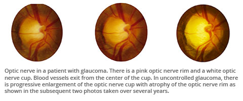
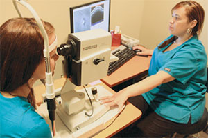 During your eye exam, our physicians carefully examine the health of your optic nerve. Shaped liked a doughnut, the optic nerve has a round rim of neural fibers (pink) and often a central area lacking such tissue (white). In the photos below, note that as glaucoma progresses from mild to advanced stages, the pink areas of the nerve rim become more narrow or thin while the white area of the nerve (the “cup”) expands. For many patients, pupil dilation provides the best view of your optic nerve. Optic nerve scanning, shown in the photo to the right, is performed in the office to determine if damage to nerve fibers has occurred.
How is visual field testing performed?
Routine eye exams are the best way to identify glaucoma in its early stages and before significant vision loss has occurred. Glaucoma results in optic nerve damage and subsequent vision loss is not reversible. However, glaucoma can be controlled with treatment and blindness can be prevented.
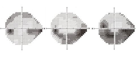
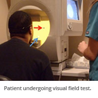 In this exam, the patient fixates on a central target (red arrow) while white dots at varying intensities of brightness appear at varying locations in the visual field, centrally and peripherally. In this photo, a faint white dot appears in the lower portion of the patient’s visual field. When the patient detects the white dot of light, he then pushes a button indicating he saw the dot of light. The technician monitors the patient’s fixation and responses and the computer analyzer produces results similar to the figures above. In the pictured visual field, the central cross hairs are where the patient is fixating. The dark spot towards the left is the normal blind spot that everyone has with their left eye.
In the far left figure of the series (below), a patient with mild glaucoma was not able to perceive the white dot stimuli towards his lower right side, identified by dark areas. As glaucoma progresses, the dark areas enlarge to deplete most of his lower vision permanently. In most situations, patients are completely unaware that they have lost this much of their vision. Hence, visual field testing is an essential test to identify glaucomatous visual field loss and progression before the patient becomes aware of it. By the time a patient notices that he or she is losing vision, it is often too late and the lost vision cannot be recovered.
How is glaucoma treated?
Treatment for glaucoma is targeted at lowering pressure inside the eye, which is the most common cause for optic nerve damage. Pressure control is usually achieved with medication. When medication is insufficient in preventing nerve damage or vision loss, laser therapy or surgery is the next step of therapy.
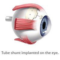
Traditional glaucoma surgery includes trabeculectomy and tube shunt implantation.
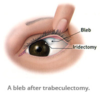 A trabeculectomy lowers the pressure inside the eye by making a small hole in the eye. The fluid inside the eye drains to a small reservoir or bleb just under the eye surface, hidden by the upper eyelid.
A tube shunt consists of a small silicone tube attached to a plate. The tube drains the fluid from inside the eye to the plate which sits on the white of the eye behind the eyelid.
All of the eye physicians at University Eye Specialists are experts in glaucoma care, each having completed a fully accredited glaucoma fellowship. We are a tertiary practice for complex glaucoma cases referred from eye care professionals throughout Chicagoland, as well as from physicians around the country.
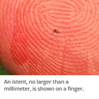 Among treatments for mild to moderate glaucoma, the newest surgical technology is Minimally Invasive Glaucoma Surgery (MIGS). MIGS targets the natural drainage system of the eye found in the “angle” between the iris and the cornea. The goal of MIGS is to reduce your eye pressure with less risk, a shortened recovery time, and fewer follow up visits. MIGS includes goniotomy, ab-interno canaloplasty, gonio- assisted transluminal trabeculotomy, iStent, and others.
University Eye Specialists performed the first iStent procedures in Chicago. Watch a video about iStent glaucoma treatment »
Learn more about Minimally Invasive Glaucoma Surgery and the types of MIGS options that our surgeons offer. »
Learn how University Eye Specialists distinguishes itself in the management of glaucoma patients »
Back to top